Medical & Research
Case Studies
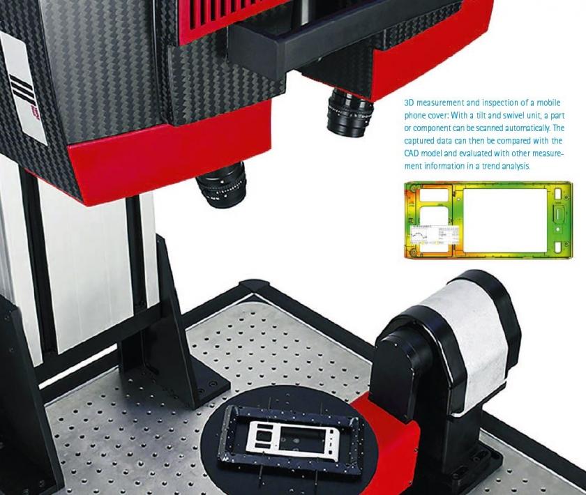
Learn how medical and research companies are using accurate structured blue light 3D scanners, comprehensive inspection software, photogrammetry technology, and/or automation to improve manufacturing processes with rapid precision measurements

This application shows the complete digitizing of the model of a torso and its removable organs, with all data in one coordinate system, using the…
Due to the technological developments of recent years, the institute of forensic medicine of the University of Bern, Switzerland, and the scientific service of the…
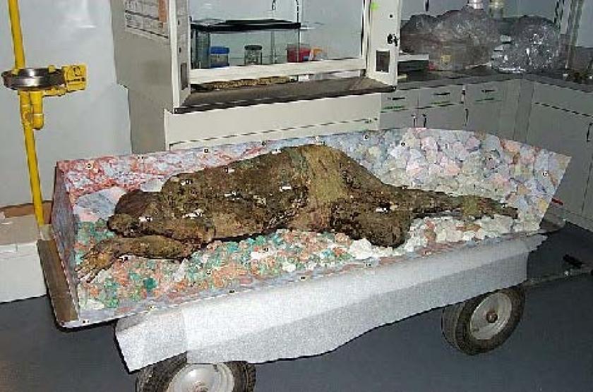
Scansite was contracted to recreate a Chilean mummy with ATOS optical non-contact 3D scanner. This presentation also gives an overview of their other art and…
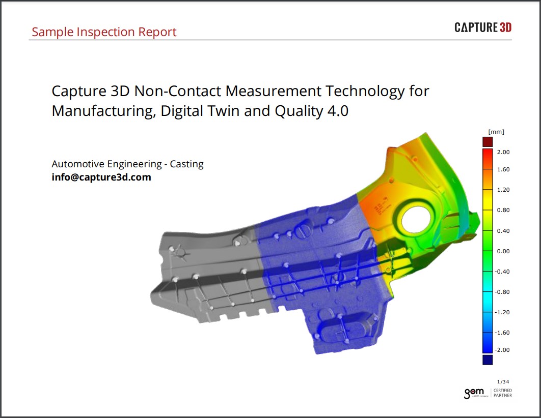
In recent years, the complexity of parts in the plastics processing industry has increased, generating high demand for the development and use of new metrology…
Orchid Orthopedic Solutions is a medical device manufacturer providing contract manufacturing services for orthopedic procedures to original equipment manufacturers (OEMs) and other companies within the…
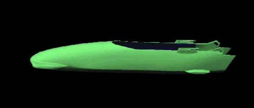
Sammer Technologies was contracted in summer 2007 to provide 3D models of 4 man and 2 man bobsleds with crews.
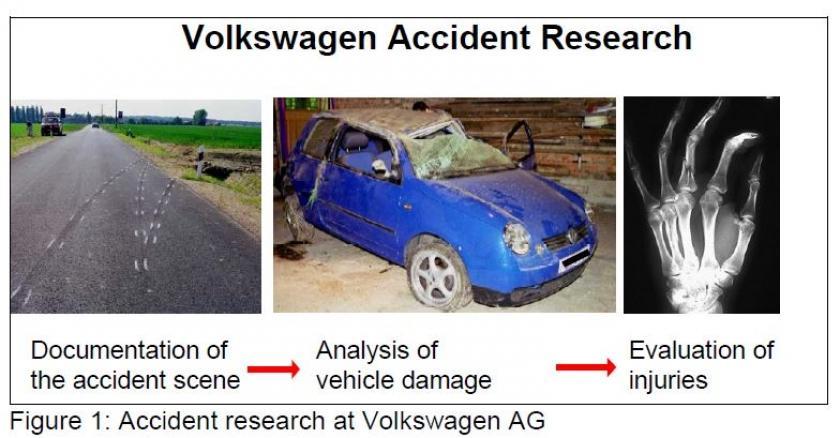
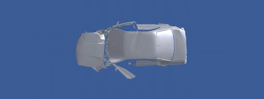
Volkswagen pursues accident research. Numerous accident statistics and local accident surveys have been analyzed statistically to gain knowledge of the accident event. The goal is…